5.1.2 The torso and pelvis segments
Ergoniza Intelligent models the trunk using two segments: the torso and the pelvis. The torso segment comprises the thorax and abdomen and extends from the L5/S1 lumbosacral joint to the C7/T1 region. The pelvis segment extends from L5/S1 to the hip joints.
The trunk has three degrees of freedom: flexion and extension, left and right lateral flexion, and left and right rotation. Lateral-flexion and rotation movements are performed by the torso segment through its rotation relative to the pelvis at L5/S1, while the pelvis remains fixed. In contrast, trunk flexion and extension are distributed between the torso and pelvis segments. This distribution is explained in the following section.
5.1.2.1 Modeling flexion at the lumbosacral joint
In the Ergoniza Intelligent model, total trunk flexion results from the combination of two movements: rotation of the pelvis relative to the thighs at the hip joints and flexion of the torso relative to the pelvis in the lumbosacral region. Several studies have shown that the thoracic vertebrae exhibit little relative movement during trunk flexion and that a substantial part of the movement occurs in the lumbar region1.
Flexion from an upright position begins with rotation of the lumbar vertebrae. In general, as trunk flexion increases, the pelvis begins to rotate together with the spine, although at a different rate. The rotation rates assumed by the spine and pelvis have been analyzed in different studies to obtain a quantitative model of spinal kinematics during trunk flexion2. These studies have shown that the degree of sacral rotation at the hip joints and the rotation of the L5 vertebra relative to the sacrum depend not only on the degree of trunk flexion but also on the knee-flexion angle. Other factors, such as the load handled in the hands, sex, body weight, or somatotype, appear to have a smaller effect on the lumbosacral-joint rotation pattern3.
Ergoniza Intelligent models the relative movements between the torso and pelvis segments during trunk flexion and extension using these kinematic models. At the beginning of flexion from an upright position, most rotation occurs in the lumbar region. As the trunk-flexion angle increases, the rate of sacral rotation at the hips increases until it predominates over rotation in the lumbar region. The effect of knee flexion is also considered: as knee flexion increases, the relative amount of sacral rotation decreases, while relative rotation at L5/S1 increases.
An example is shown in Figure 7. The postures in the figure have the same trunk flexion, while knee flexion is greater in posture B. The distribution of flexion between the pelvis and torso differs between the two postures. Sacral rotation is lower in posture B because knee flexion is greater.
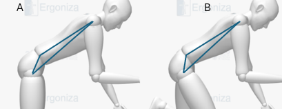
Figure 7: Variation in the relative rotation rate between the pelvis and torso with the degree of knee flexion.
5.1.2.2 Modeling the trunk musculature
Ergoniza Intelligent estimates compression and shear forces at the L4/L5 intervertebral disc. The magnitude of these forces and their comparison with the corresponding reference values provide relevant information for assessing tasks that may generate high loads in the lumbar spine.4
To solve moment equilibrium at L4/L5, the model incorporates ten muscle units distributed across five bilateral pairs: latissimus dorsi, erector spinae, rectus abdominis, obliquus externus, and obliquus internus. For each unit, the model considers its line of action, its moment arm relative to the spine, and its cross-sectional area at L4/L55. The calculation also incorporates the effect of intra-abdominal pressure.
The procedure used to determine muscle forces and calculate compression and shear forces at L4/L5 is described in detail in Section 12.
Peter R. Davis, “Posture of the Trunk during the Lifting of Weights,” British Medical Journal, 1959, doi:10.1136/bmj.1.5114.87; S. Kumar, “A Study of Spinal Motion during Lifting,” Irish Journal of Medical Science 143, no. 2 (1974): 86–95, doi:10.1007/BF03004745; Elizabeth A. Tully, Mohammad Reza Fotoohabadi, and Mary P. Galea, “Sagittal Spine and Lower Limb Movement during Sit-to-Stand in Healthy Young Subjects,” Gait and Posture, 2005, doi:10.1016/j.gaitpost.2004.11.007; P. R. Davis, J. D. Troup, and J. H. Burnard, “Movements of the Thoracic and Lumbar Spine When Lifting: A Chrono-Cyclophotographic Study,” Journal of Anatomy 99 (1965): 13–26.↩︎
C. K. Anderson, D. B. Chaffin, and G. D. Herrin, “A Study of Lumbosacral Orientation under Varied Static Loads,” Spine 11, no. 5 (1986): 456–62, doi:10.1097/00007632-198606000-00012; C K. Anderson et al., “A Biomechanical Model of the Lumbosacral Joint during Lifting Activities,” Journal of Biomechanics 18, no. 8 (1985): 571–84, doi:10.1016/0021-9290(85)90012-0.↩︎
D. Allbrook, “Movements of the Lumbar Spinal Column.,” The Journal of Bone and Joint Surgery. British Volume, 1957, doi:10.1302/0301-620x.39b2.339.↩︎
T R Waters et al., “Revised NIOSH Equation for the Design and Evaluation of Manual Lifting Tasks.,” Ergonomics 36 (1993): 749–76, doi:10.1080/00140139308967940.↩︎
S. McGill, Low Back Disorders. Evidence-Based Prevention and Rehabilitation (Champaign, Illinois: Human Kinetics, 2007); D. B. Chaffin et al., “Lumbar Muscle Size and Locations from CT Scans of 96 Women of Age 40 to 63 Years,” Clinical Biomechanics 5, no. 1 (1990): 9–16, doi:10.1016/0268-0033(90)90026-3.↩︎